Cavovarus Foot Correction
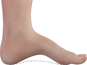
To support the entire body's weight on your two feet, the inner middle portion of each foot (midfoot) is raised off the ground to form an arch. A cavovarus foot deformity is characterized by a higher-than-normal arch of the inner midfoot. This results as the two ends of the foot - the heel and toes - abnormally draw towards the inside of the foot, causing the foot to rest on its outer side. This deformity produces pain in your heel, ball of the foot and outer edge of the foot, instability of gait, frequent ankle sprains, difficulty wearing shoes, callus formation and sometimes stress fractures in the bones on the outer side of the foot.
The cause for cavovarus foot deformity is usually unknown, but it may be associated with neuromuscular conditions such as Charcot-Marie-Tooth disease (progressive muscle weakness), stroke, head injury and poliomyelitis (viral infection that causes paralysis). It may be produced by an imbalance in the strength of the foot muscles, causing muscle contractures (stiffness) or due to bony deformities of the heel bone.
Cavovarus deformity may be corrected by conservative methods, such as bracing, to help with ankle instability and sprains, and shoe inserts, to raise the lateral border of the foot and accommodate the middle region of the foot. If cavovarus deformity is not adequately controlled by conservative means, your doctor will recommend surgical treatment. Weak muscles and contractures are corrected by a tendon transfer surgery, while bone deformities are corrected by cutting (osteotomy) or fusing bones (arthrodesis) to allow the foot to evenly contact the floor.